Keratoconus
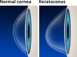
 The cornea is the clear front window of the eye. Normally, the cornea is shaped like a dome. Patients with keratoconus have corneas that are steeper and thinner than normal.
These changes to the cornea first begin in the teenage years and then progress at an unpredictable rate during a patient’s 20s and 30s. Typically, the cornea will stop changing sometime during a patient’s 40s.
Initially, it may be possible to correct the vision of a keratoconus patient using spectacles or soft contact lenses. However, as the changes to the shape of the cornea become more severe, patients can achieve their best vision only with the use of a hard contact lens. Most keratoconus patients eventually require hard contact lenses, but most are able to enjoy good vision throughout their lives without any further treatment.
For some patients, the cornea becomes so protuberant that contact lenses no longer fit and/or scarring occurs. In these cases, a patient’s best vision can only be achieved through surgery (a corneal transplant).
If keratoconus is diagnosed at an early enough age, crosslinking can be performed to stabilize the cornea. This procedure reduces the risk of future surgery, and it increases the likelihood of maintaining good vision with the use of only spectacles or soft contact lenses.
Visit the American Academy of Ophthalmology’s EyeSmart® website to learn more about Keratoconus.


